7.1
Life is Cellular
Can we just keep dividing a living organism in half and it still be alive?
There is a limit. The smallest living unit of any organism is the cell.
As is usual, discoveries in one science lead to new discoveries in other sciences. With the invention of the microscope, we were able to then discover that living things were made of cells.
Robert Hooke used an early compound microscope to look at a nonliving thin slice of cork. He called the chambers "cells" because they resembled the cells that monks lived in.

Anton van Leeuwenhoek used a single-lens microscope to observe pond water. The microscope revealed many tiny creatures previously unseen to the naked eye. He drew pictures of his findings.

The work of three men, Schleiden, Schwann, and Virchow, was eventually tested and confirmed by many biologists and became known at the Cell Theory.
The Cell Theory
1. All living things are made up of cells
2. Cells are the basic units of structure and function in living things
3. New cells are produced from existing cells
Most microscope focus light or electrons on an object to magnify an image.
Light Microscopes
A typical light microscope allows light to pass through a specimen and uses two lenses to form an image. The first lens, called the objective lens, is located just above the specimen. The second lens, the ocular lens, magnifies this image still further.

Light microscopes can produce clear images of objects only to a magnification of about 1000X.
Because cells are transparent, we use chemical stains or dyes to reveal certain compounds or structures in the cell.
Fluorescent dyes can be attached to specific molecules and can then be made visible using a special fluorescence microscope.

Electron Microscopes
Electron microscopes use beams of electrons that are focused by magnetic fields. They offer much higher resolution than light microscopes.
Electron microscopes use beams of electrons that are focused by magnetic fields. Some electron microscopes can offer resolution up to 1 billionth of a meter in size.
Two type of electron microscopes:
Scanning Electron Microscopes (SEM) - a pencil-like beam of electrons is scanned over the surface of a specimen. SEMs allow a 3D image to be viewed.

Transmission Electron Microscope (TEM) - make it possible to explore cell structures and large protein molecules. The beams of electrons can only pass through thin samples, cells and tissues must be cut into ultrathin slices before they can be examined. For this reason, images are 2D.

 |
| Add caption |
|
| Add caption |
 |
| TEM Prokaryotes and Eukaryotes Cells come in many shapes and sizes. Typical cells range from 5-10 micrometers in diameter. The smallest Mycoplasma bacteria are 0.2 micrometers across. The giant one-celled amoeba Chaos chaos is up to 1 mm in diameter, and can be seen as a tiny speck in pond water.  All cells at some point in their lives contain DNA. All cells are surrounded by a thin flexible barrier called a cell membrane. Cells fall into two categories: Eukaryotes - DNA is enclosed in a membranes bound nucleus Prokaryotes - DNA is not enclosed in a membrane bound nucleus The simplest organisms (bacteria) are prokaryotic. The more complex organisms (plants and animals) are eukaryotic. For Powerpoint 7.1 click here 7.2 Cell Structure  Eukaryotic cells have many structures, each with an important role in the cell and the organism as a whole. These structures are called organelles, literally meaning "little organs". Cytoplasm (Cell juice) -a jelly-like substance that is the portion of the cell outside the nucleus where the other organelles float. The nucleus contains nearly all the cell's DNA and, with it, the coded instructions for making proteins and other important molecules. The DNA is the hereditary material which is passed from generation to generation of cells.   The nucleus is surrounded by a nuclear envelope composed of two membranes. The envelope is dotted with thousands of nuclear pores, which allow material to more into and out of the nucleus. A steady stream of proteins, RNA, and other molecules move through the nuclear pores to and from the rest of the cell. Chromatin, uncoiled chromosomes, a complex of DNA bound to proteins, is found in the nucleus. Most nuclei contain a small dense region known as the nucleolus, where the assembly of ribosomes begins. Storage, Clean Up, and Support Organelles Vacuoles are large, saclike, membrane-enclosed structures which store materials like water, salts, proteins, and carbohydrates.  Plants tend to have one main large vacuole called a central vacuole which stores much of the water in a plant.  Paramecium have a contractile vacuole, which pumps excess water out of the cell, propelling them through water. 
Lysosomes are small organelles filled with enzymes. They break down lipids, carbohydrates, and proteins into small molecules that can be used by the rest of the cell. They are also involved in breaking down organelles that have outlived their usefulness. A number of serious human diseases can be traced to lysosomes that fail to function properly.

The cytoskeleton gives eukaryotic cells their shape and internal organization by a network of protein filaments. It also helps provide movement for the cell. Two kinds of proteins make up part of the cytoskeleton:
Microfilaments are threadlike structures made up of a protein called actin. They form extensive networks in some cells and produce a tough flexible framework.
 
Microtubules are hollow structures made up of proteins known as tubulins. These play a critical role in cell shape. They are also important in cell division where they form the mitotic spindle. In animal cells, they also form structures call centrioles which are located near the nucleus and help organize cell division.
Microtubules help build projections from the cell surface known as cilia and flagella. In these structures, microtubules are lined up in a 9 + 2 formation producing controlled movements.

Organelles that Build Proteins
Living things are always working, building new molecules all the time, especially proteins which have multiple uses.
Proteins are assembled on ribosomes. Ribosomes are small particles of RNA and protein found throughout the cytoplasm in all cells. They produce proteins by following coded instructions that come from DNA.

Smooth ER is a site of cell detoxification and synthesis of membrane lipids.
Endoplasmic Reticulum is an internal membrane system. The "ER" is where lipid components of the cell membrane are assembled, along with proteins and other materials that are exported from the cell.
The Rough ER is involved in the synthesis of proteins. It is called "rough" because that is where the ribosomes are found. Newly made proteins leave these ribosomes and are inserted into the rough ER, where they may be chemically modified.

Proteins made on the rough ER include those that will be released, or secreted, from the cell as well as many membrane proteins and proteins destined for lysosomes and other specialized location within the cell. Rough ER is abundant in cells that produce large amounts of protein for export.
Proteins then travel to the Golgi Apparatus, a stack of flattened membranes, where they are modified, sorted, and packaged according to the instructions sent with them from the DNA/RNA. They may leave the Golgi inside vesicles and go live inside the cell, outside the cell, or in the cell membrane.

Organelles that Capture and Release Energy
Cloroplasts are the bio equivalents of solar power plants. Chloroplasts capture energy from sunlight and convert it into food that contains chemical energy in a process called photosynthesis.
 Mitochondria are the power plants of the cell. They convert chemical energy stored in food into compounds that are more convenient for the cell to use (ATP). All mitochondria come from the mother. 
Cellular Boundaries
Cell walls are found in plants and prokaryotes (bacteria) and they provide strong support for the cell while allowing gases and water needed for photosynthesis to pass through.

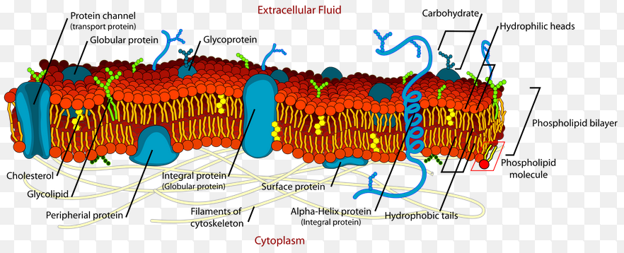
All cells have cell membranes. The membrane is composed of a lipid bilayer with transport proteins and carbohydrate chains imbedded in it. The phospholipid has a water-loving (hydrophilic) head and water-hating (hydrophobic) tails.
The membrane is referred to as the Fluid Mosaic Model and is selectively permeable, allowing only certain substances to pass through.

7.3 Cell Transport
The cell membrane is a selectively permeable membrane, meaning that some substance can pass across them and others cannot.
Passive Transport
Passive transport is the movement of materials across the cell membrane without using cellular energy. There are 3 types of passive transport:
1. Diffusion
In any solution, solute particles move constantly. They collide with one another and tend to spread out randomly. As a result, the particles tend to move from an area where they are more concentrated to an area where they are less concentrated. This process is called diffusion. Diffusion, which does not require energy, is the driving force for many substances moving across the cell membrane.
A net movement from an area of high concentration to an area of lower concentration will continue until the concentrations of solute on both sides of the membrane are equal, or equilibrium is reached.

2. Facilitated Diffusion
Sometimes molecules are too large to pass through the lipid bilayer, or they are not soluble in the lipid layer, and must be given a different and larger pathway. Some proteins embedded in the membrane have channels for these larger molecules to pass through. These "channel proteins" allow larger substances to diffuse through the membrane through diffusion. The proteins help the substance to pass through, thereby, facilitating the process. This process is known as facilitated diffusion.
 
Aquaporins are proteins that are specific for facilitating water to diffuse through a cell membrane.
3. Osmosis
Osmosis is the diffusion of water through a selectively permeable membrane. If a solute cannot move through a cell membrane, then water will move through aquaporins to create the equilibrium.
 
When two solutions on either side of a membrane are the same solute strength, the solution is said to be isotonic.
When the solution outside a cell has a higher solute concentration than inside, the solution is said to be hypertonic.
When the solution outside the cell has a lower solute concentration than inside, the solution is said to be hypotonic.

Water molecules move equally into and out of cells placed in an isotonic solution. In a hypertonic solution, animal cells, like the red blood cell shown, shrink, and plant cell central vacuoles collapse. In a hypotonic solution, animal cells swell and burst. The central vacuoles of plant cells also swell, pushing the cell contents out against the cell wall.
Osmotic Pressure is the force produced by the net movement of water out of or into a cell driven by differences in solute concentration.
Organisms like fish and amphibians that lay eggs in fresh water have cells that lack water channels so the eggs don't expand too much. Other organisms like bacteria and plant cells have cell walls that prevent the cells from expanding, even under tremendous osmotic pressure.
Active Transport
1. Molecular Transport
In active transport, cells must move materials against a concentration difference, or from high to low concentration. Active transport requires energy. Active transport of small molecules or ions across a cell membrane is generally carried out by transport proteins, called protein pumps, found in the membrane. These pumps use energy to move calcium, potassium, and sodium across cell membranes. This is necessary to create a muscle contraction or to send a nerve impulse. A considerable amount of energy is used in this process.

2. Bulk Transport
Larger molecules and even solid clumps of material can be transported by movements of the cell membrane known as bulk transport.
Endocytosis is the process of taking material into the cell by means of infoldings, or pockets of the cell membrane.
The pocket that results breaks loose from the outer portion of the cell membrane and forms a vesicle or vacuole within the cytoplasm. Large molecules, clumps of food, even whole cells can be taken up in this way.
The white blood cell seen here is engulfing a damaged red blood cell by phagocytosis.
Two specific types of endocytosis are
phagocytosis - cell eating
and pinocytosis - cell drinking
Exocytosis releases large amounts of material in bulk. The membrane of a vacuole surrounds the material and fuses with the cell membrane, forcing the contents out of the cell.
 |
7.4 Homeostasis and Cells
In order to survive, cells and organisms must maintain homeostasis, constant internal physical and chemical conditions. To maintain homeostasis, unicellular organisms grow, respond to the environment, transform energy, and reproduce.

Homeostasis is an issue for prokaryotic organisms because if the cell becomes unstable and dies, the organism dies. In eukarytotic organisms, one cell death will not normally affect the whole organism adversely.
The cells of a multicellular organism rely on each other like the members of a baseball team. Each cell has become a specialist at it's job and is relied on by other cells and, vice versa. The cells of a multicellular organism become specialized for particular tasks and communicate with one another to maintain homeostasis.
Specialized Animal Cells

Some human cells specialize to be cells which line the airways leading to the lungs. Since so much debris is entering the lungs all the time the body has to fight back to prevent particles in the air from building up in the lung aveoli where gas exchange takes place. One way this happens is due to the specialized cells which have cilia constantly pumping the debris caught in the mucus back up toward the opening it came in.

The cell is the smallest living unit. Specialized cells of multicellular organisms are organized into tissues, then into organs, and finally into organ systems, which make up the organism.

Cellular Communication
In multicellular organisms, cell communication is a must. Cells in large organisms communicate by means of chemical signals that are passed from one cell to another.
To respond to these chemical signals, the cells have receptors to which the signaling molecule can bind. The signal will tell the specialized cell to either speed up or slow down its job.
Some cells communicate through junctions between the cells seen below.

Gap junctions are connections between heart muscle cells so that they can communicate and contract in sync.
For Powerpoint 7.4 Click here

Plant sperm cells (pollen) have specialized with "wings" which help the pollen fly through the air, helping to spread it out, and increasing the chance of fertilization.
Levels of Organization
The cell is the smallest living unit. Specialized cells of multicellular organisms are organized into tissues, then into organs, and finally into organ systems, which make up the organism.

Cellular Communication
In multicellular organisms, cell communication is a must. Cells in large organisms communicate by means of chemical signals that are passed from one cell to another.
To respond to these chemical signals, the cells have receptors to which the signaling molecule can bind. The signal will tell the specialized cell to either speed up or slow down its job.
Some cells communicate through junctions between the cells seen below.
Gap junctions are connections between heart muscle cells so that they can communicate and contract in sync.
For Powerpoint 7.4 Click here


