Sunday, April 26, 2015

Ch 12 DNA

12.2 The Structure of DNA


There were several people who are credited with finding the structure of this most important molecule.

Remember the 4 organic molecules (molecules that make up living things)? 
carbohydrates
lipids
proteins
nucleic acids

DNA is a nucleic acid made up of nucleotides joined into long strands or chains by covalent bonds.

Nucleic Acids are long, slightly acidic (hence, the name) molecules usually found in the cells nucleus (if there is one). They are polymers made up of smaller units (monomers) called Nucleotides.

A nucleotide contains 3 smaller molecules:
a phosphate group
a 5 carbon sugar (carb) called deoxyribose
a nitrogen base

There are 4 different types of nucleotides. The nitrogen bases can change but the sugar and phosphate stay the same.

The four nitrogen bases are:
Guanine
Cytosine
Thymine
Note: G and A are larger, 2 ring structures while C and T are smaller, single ring structures. Each single ring structure always pairs with a 2 ring structure in a double helix. An easy way to remember which are double ringed is that they are G and A, like GA (Georgia) which is a larger state than CT (Connecticut), C and T.

Nucleotides are held together to build nucleic acids by covalent bonding between the sugars and the phosphate groups along the outside of the "ladder".

The nucleotides can be joined in any order, meaning any sequence of bases is possible. 

It just so happens that this chemical structure is especially good at absorbing UV Light. This isn't necessarily a good thing because this can cause mutations in the order of nucleotides.


Chargaff's Rule

Edwin Chargaff, an Austrian-American biochemist, had discovered that the percentages of adenine (A) and thymine (T) bases are almost always equal in any sample of DNA. The same rings true for guanine (G) and cytosine (C). This observation known as "Chargaff's rule" led us to the knowledge that  T always pairs with A and G always pairs with C.


Rosalind Franklin

In the 1950's, British scientist Rosalind Franklin began to study DNA. Using x-ray diffraction, she was able to record the scattering pattern of DNA shows that the strands in DNA are twisted around each other like the coils of a spring, a shape known as a helix.

Watson and Crick
American biologist, James Watson, and British physicist, Francis Crick, had built 3D models of the molecule... but not until in 1953, when they saw Franklin's remarkable X-ray pattern, did they figure out the correct double helical structure for DNA.


Watson and Crick's model of DNA was a double helix, in which two strands of nucleotide sequences were wound around each other. This explains Chargaff's rule of base pairing and how the two strands of DNA are held together.


Antiparallel Strands
The two strands of connected DNA run in opposite directions, and are "anti-parallel".
The two strands are joined at the nitrogen bases and are held together by Hydrogen Bonds. H bonds are weaker bonds than covalent. This is helpful when the strands have to separate for DNA replication in cell division.
Note that C and G are always held by 3 H bonds, while A and T are always held by 2 H bonds.
This nearly perfect fit between A-T and G-C nucleotides is known as base pairing.


Watch Hank's great description here about the structure and replication of DNA!
                                        

For 12.2 Powerpoint click here:


12.3 DNA Replication


Base pairing in the double helix explains how DNA can be copied!

Each strand of the double helix has all the info needed to reconstruct the other half by using base pairing. The strands are said to be complementary.

Before a cell divides, it duplicates its DNA in a copying process called replication, which occurs during the S phase of interphase. During replication, the DNA molecule separates into two strands and then produces two new complementary strands. Each original strand serves as a template, or model, for the new strand.

During replication, the two original strands of the double helix "unzip", allowing two replication forks to form. Each strand now becomes a template for its complimentary new strand to form on. Each of the two new double strands are made up of one original strand and one new strand. 

Enzymes

DNA replication is carried out by a series of enzymes. 

1. Enzymes "unzip" a molecule of DNA by breaking the H bonds between the base pairs and unwinding the two strands of the molecule.

2. DNA polymerase joins individual nucleotides to produce a new strand of DNA. Then, DNA polymerase adds the sugar-phosphate bonds that hold the nucleotides together. Finally, DNA polymerase also "proofreads" each new strand to check for mistakes.


Micrograph showing a pair of replication forks in human DNA

Telomeres
Telemeres are the white (stained) part of the blue human chromosome
DNA at the tips of chromosomes are known as telomeres. These telomeres sometimes like to break off. This DNA is particularly difficult to replicate. Cells use a special enzyme, telomerase, to solve the problem by adding short, repeated DNA sequences to the telomeres.

Prokaryotic DNA Replication

Remember that in prokaryotes, there is no nucleus to contain the DNA. Prokaryotes have a single, circular DNA molecule in the cytoplasm, containing nearly all the cell's genetic info. 

DNA replication begins when a regulatory protein binds to a single starting point on the chromosome. These proteins trigger the beginning of the S phase, and DNA replication begins.


Replication in most prokaryotic cells starts from a single point and proceeds in two directions until the entire chromosome is copied. The two chromosomes produced by replication are attached to different points inside the cell membrane and are separated when the cell splits.

Eukaryotic DNA replication

Eukaryotic chromosomes are generally much bigger than those of prokaryotes. In eukaryotic cells, replication may begin at dozens or even hundreds of places on the DNA molecule, proceeding in both directions until each chromosome is completely copied.


The system is not foolproof even though a number of proteins check DNA for chemical damage or base pair mismatches prior to replication. 

The two copies of DNA produced by replication in each chromosome him remain closely associated until the cell enters prophase of mitosis. At that point, the chromosomes condense, and the two chromatids in each chromosome become visible.


What this video to help understand DNA replication!

Ba
For 12.3 Powerpoint click here:


Sunday, March 15, 2015

Ch 11 Intro to Genetics

Chapter 11 Genetics


11.1 The Work of Gregor Mendel


What's your inheritance?
The modern science of genetics was founded by an Austrian monk named Gregor Mendel. Mendel spent several years studying science and started genetics experiments in the monastery gardens on pea plants. 


He used ordinary garden peas because they are small and easy to grow. Also, they produces hundreds of offspring in a short life cycle. Today, his research with pea plants is called a model system.

Fertilization
During sexual reproduction, male and female reproductive cells join in a process known as fertilization to produce a new cell (organism). The new cell develops into a tiny embryo encased within a seed. 
Flowers are the reproductive organs on plants. Pea flowers are normally self-pollinating, which means that sperm cells fertilize egg cells from within the sam flower. A plant grown from a seed produced by self-pollination inherits all of its characteristics from the single plant that bore it; It has a single parent. Therefore, the traits of each successive generation would be the same. Traits are specific characteristics of an individual. 
Cross-Pollination
Mendel decided to cross-pollinate the pea flowers by removing the male reproductive organs from one plants flower and fertilizing the female reproductive organ on another plant. In this way, the new organism will have two parents with different DNA. By crossing two plants with different traits, he was able to study how genes are passed and expressed over generations. Crossing two organisms with different traits creates a hybrid

Mendel studied seven different traits of pea plants. 
When doing genetic crosses, the original pair of plants is called the P or parental generation. Their offspring are called the F1 or first filial generation. The grandchildren of the P generation are the F2 generation.

Much to his surprise, when Mendel crossed two plants with different traits, only one of the traits showed up in the offspring. Where did the other one go?

Mendel drew two conclusions:

1. An individual's characteristics are determined by factors that are passed from one parental generation to the next. Today we call these factors genes. Different forms of a gene are called alleles. For example, everyone has a gene for eye color. The different alleles for eye color might be blue, brown, green, and hazel.

2. Principle of Dominance: states that some alleles are dominant and others are recessive. An organism with a least one dominant allele for a particular form of a trait will exhibit that form of the trait. The recessive allele for a particular form of a trait will exhibit that form only when the dominant allele for the trait is not present.


Mendel discovered that the round seed shape, yellow seed color, gray seed coat, smooth pod shape, green pod color, axial flower position, and tall plant height were the dominant alleles for pea plants.


If recessive alleles disappear in the 2nd generation, where did they go?

Mendel continued to breed the 2nd generation with itself by self-pollination and found that the recessive traits showed up again in the 3rd generation. 

Mendel assumed that a dominant allele had masked the corresponding recessive allele in the F1 generation. However, because the recessive trait showed up in the F2 generation. This showed that the F1 plants carried the recessive allele.

Segregation
During the production of gametes (sex cells), eggs and sperm, reproductive organs undergo meiosis. Meiosis produces cells with half the normal number of chromosomes. Therefore, the sperm and egg cells only carry one allele for  each genes. This is a product of meiosis, or segregation:

After each parent cell segregates and forms gametes, the offspring could inherit either of the two parent's alleles, strictly by chance of which gamete it formed from. 


For 11.1 powerpoint please click link below:

https://docs.google.com/presentation/d/1z57BO3TQKotC62yP5b8kgyRSz6jg_8j7aBisEnEA1Ts/edit?usp=sharing

11.2 Applying Mendel's Principles

Probability

Probability is the chance that an event or outcome will occur. Mendel uses probability to describe the different probable outcomes of offspring after cross-pollinating.

In a coin flip, there are two sides to a coin so there are two possible outcomes. The probability that the coin will land on one of the two sides is 1/2, or 50%.

In segregation, each of the two parent alleles separate to form gametes or sex cells. The gametes then only carry one of the parents alleles. The parent has a 50-50% chance of passing on either allele to their offspring. This happens purely by chance. The offspring could inherit either allele from the parent. The new offspring will inherit only one allele from one parent and only one allele from the other parent so that the new offspring now has two of its own alleles.


Homozygous and Heterozygous

Homozygous - organisms that have two identical alleles for the same gene

Heterozygous - organisms that have two different alleles for the same gene

Eye color

Homozygous

BB (homozygous dominent)
or
bb (homozygous recessive)

Heterozygous

Bb (the dominant allele will show up)

Probabilities Predict Averages

Probabilities can predict the average outcome of a large number of events. If you flip a coin twice, you could get one heads and one tails. Or, you could get two heads or two tails. 

The larger the number of coin flips, or offspring, the closer the results will be to the predicted values.

Genotype and Phenotype

Every organism has a genetic makeup as well as a set of observable traits.

The genotype is the genetic makeup of an organism for any given gene. It contains two alleles for that gene. For instance, a persons genotype for eye color could be Bb. (Geno means "race, kind")

The phenotype is the observable characteristic of the trait. For instance, the phenotype for eye color for a person with Bb would be Brown (even though they also carry a blue gene). (Pheno means "to show")

The genotype can be homozygous dominant, homozygous recessive, or heterozygous.

The phenotype will be only the dominant allele or the recessive allele.

Two organisms can have the same phenotypes but different genotypes.

PUNNETT SQUARES

Punnett Squares use mathematical probability to help predict the genotype combinations in genetic crosses. This diagram is one of the best ways to predict the outcome of a genetic cross.


Using the Punnett Square you can deduce that the offspring will have one of the four above genotypes, BB, Bb, bB, or bb. There are actually only three possibilities because Bb and bB are the same genotype. From these genotypes you can deduce that the phenotypes may be dominant or recessive. In this case the probabilities of having a dominant phenotype is 3/4 or 75% and a recessive phenotype is 1/4 or 25%.


It is possible to determine the probability when two or more factors are involved as well.

 In this example, the color of the squares represents pod color. Alleles in black indicate short plants, while alleles in red indicate tall plants


Mendel crossed plants that were homozygous dominant for round yellow peas with plants that were homozygous recessive for wrinkled green peas below.

All of the F1 offspring were heterozygous dominant for round yellow peas.

Below, when Mendel crossed F1 plants that were heterozygous dominant for round yellow peas...
He found that the alleles segregated independently to produce the F2 generation.

Independent Assortment
The principle of independent assortment states that genes for different traits can segregate independently during the formation of gametes. Independent assortment helps account for the many genetic variations observed in plants, animals, and other organisms--- even when they have the same parents!


Summary of Mendel's Principles

Mendel's principles of heredity, observed through patterns of inheritance, form the basis of modern genetics.

1. The inheritance of biological characteristics is determined by individual units called genes, which are passed from parents to offspring.
2. Where two or more forms (alleles) of the gene for a single trait exist, some alleles may be dominant and others may be recessive.
3. In most sexually reproducing organisms, each adult has two copies of each gene---one from each parent. These genes segregate from each other when gametes are formed.

4. Alleles for different genes usually segregate independently of each other.

Mendel's principles apply to all organisms that reproduce sexually. In the early 1900s, Thomas Hunt Morgan used the insect known as the fruit fly, Drosophila melanogaster, to reproduce the same results that Mendel gave. A single pair of fruit flies can produce hundreds of offspring.


For 11.2 Powerpoint click below


11.3 Other Patterns of Inheritance

Exceptions to Mendel's Rules

Sometimes, some alleles are neither dominant nor recessive.

Incomplete dominance - the heterozygous phenotype lies somewhere between the two homozygous phenotypes. Example: if a chicken has incompletely dominant alleles for black and white feathers, the feather may turn out grey. 

In four o'clock plants, the alleles for red and white flowers show incomplete dominance. Heterozygous (RW) plants have pink flowers - a mix of red and white coloring.

Codominance: the phenotypes produced by both alleles are clearly expressed. Example: in the case of chickens with a black and a white allele, rather than "mixing" the two colors, there will be areas of black and areas of white making the chicken appear speckled instead of grey (incomplete dominance).


Red blood cells carry antigens, molecules that can trigger an immune reaction, on their surfaces. Human blood type A carries an A antigen, type B has a B antigen, type AB has both antigens, and type O carries neither antigen. The gene for these antigens has three alleles; A, B, and O.
Multiple alleles: Many genes exist in several different forms and are therefore said to have multiple alleles. When there are more than two alleles for a gene, there is said to be multiple alleles.
A and B are dominant alleles. O is a recessive allele.
Therefore, a person whose phenotype for blood is A can have AA or AO genotype.
A person whose phenotype for blood is B can have BB or BO phenotype.
A person whose phenotype for blood is O has a genotype of OO, because it is recessive.
And, because A and B are codominant, a person who has a genotype of AB has a phenotype of AB.
O is the universal donor. AB is the universal recipient.


Polygenic Traits - traits that are produced by the interaction of several genes. For example, at least three genes are involved in making the reddish-brown pigment in the eyes of fruit flies. And, human skin color comes from a combination of four different genes.

Genes and the Environment
Environmental conditions can affect gene expression and influence genetically determined traits.

Temperature and Wing Color
Western white butterflies that hatch in the spring have darker wing patterns than those that hatch in summer. The dark wing color helps increase their body heat. This trait is important because the butterflies need to reach a certain temperature in order to fly. The buckeye butterflies shown below also have different wing patterns at different times of year. These butterflies are darker in the autumn than they are in the summer 
This is an example of how the environment affects the phenotype of an organism.


For 11.3 Powerpoint click below

https://docs.google.com/presentation/d/1w9s1NFW64ePkqRzUwV1LDfs8PEr9mSOw4mooyafr2kE/edit?usp=sharing

11.4 Meiosis

An organism with two parents must inherit a single copy of every gene from each parent.  Matching chromosomes from each parent are called Homologous chromosomes. This means that, in fruit flies, each of the four chromosomes from the male parent has a corresponding chromosome from the female parent. A cell that contains both sets of homologous chromosomes is said to be diploid. When an organism produces gametes, those two sets of genes must be separated so that each gamete contains just one set of genes.

Diploid Cells: Having two complete sets of inherited chromosomes, one set from each parent (two complete sets of genes). This is the number of chromosomes that are in all body cells except the ones made in reproductive organs, the sex cells (eggs and sperm). Diploid number (twice haploid) is represented by the letter "2N".

Haploid Cells: Cells that contain only a single set of chromosomes (a single set of genes). The gametes of sexually reproducing organisms are haploid. Gametes are the sex cells (cells meant for reproduction which will combine with another sex cell to make a new organism). Haploid cells are created as a product of two cell divisions (Meiosis). Haploid number is represented by the letter "N".

In fruit flies, the diploid number (2N) of chromosomes is 8. The haploid number is 4.
 Meiosis: first stage in sexual reproduction. This is the process of making gametes for reproduction. The parent cell goes through two cell divisions to produce 4 daughter cells with half the number of chromosomes as the parent cell. Each new gamete is genetically different from each other and from the parent cell it came from.
Meiosis, like Mitosis, is preceded in the cell cycle by Interphase. During Interphase, in preparation for cell division, the DNA is duplicated.


Prophase I: After interphase I, the cell begins to divide, and the chromosomes pair up. Each replicated chromosome pairs with its corresponding homologous chromosome.

This pairing forms a structure called a tetrad, which contains four chromatids. As the homologous chromosomes form tetrads, they undergo a process called crossing-over. 

First, the chromatids of the homologous chromosomes cross over one another. Then, the crossed sections of the chromatids - which contain alleles - are exchanged. Crossing-over therefore produces new combinations of alleles in the cell. 

Metaphase I: paired homologous chromosomes line up across the center of the cell. 


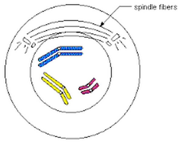
Anaphase I: spindle fibers pull each homologous chromosome pair toward opposite ends of the cell.


Telophase I and Cytokinesis: a nuclear membrane forms around each cluster of chromosomes. Cytokinesis follows telophase I, forming two new cells. 

Because of crossing over, the two cells formed from Meiosis I do not have the same exact genetic make up of each other, or the parent cell, since they "traded" some DNA in crossing-over. 

Following Meiosis I, the two new cells (2N) now enter immediately into Prophase II. Unlike before the first division, neither new daughter cell goes through a round of chromosome replication before entering meiosis II.


Prophase II: their chromosomes (each consisting of two chromatids) become visible. There are no tetrads, because the homologous pairs were already separated during meiosis I.


Metaphase II: chromosomes line up in the center of each cell.


Anaphase II: the paired chromatids separate.


Telophase II and Cytokinesis: nuclear membranes start to reform around the now haploid nuclei. Cells pinch off resulting in four haploid (N) cells (gametes). In the example (left), each of the four daughter cells produced in meiosis II received two chromosomes.


Gametes - haploid cells produced by meiosis II are gametes that are so important to heredity. In male animals, these are called sperm and all 4 sperm can go on to fertilize and egg. In female animals, these haploid cells are called eggs. Only one of the 4 female gametes will mature into an egg for reproductive purposes. 
Zygote - When a sperm fertilizes an egg, these two haploid cells will fuse to form a new diploid cell, the first cell of a new organism. The zygote will then undergo mitosis forming a new organism.

Comparing Mitosis and Meiosis

Mitosis - asexual reproduction, used for growing and repairing in multicellular organisms
Meiosis - sexual reproduction, used to produce gametes for producing new organisms


In mitosis, when the two sets of genetic material separate, each daughter cell receives one complete set of chromosomes. In meiosis, homologous chromosomes line up and then move to separate daughter cells. As a result, the two alleles for each gene are segregated, and end up in different cells. This sorting and recombination of genes in meiosis result in a greater variety of possible gene combinations than could result from mitosis.

Also, mitosis does not normally change the chromosome number of the original cell. This is not the case for meiosis, which reduces the chromosome number by half.

And finally, mitosis is a single cell division, resulting in the production of two identical daughter cells. On the other hand, meiosis requires two rounds of cell division, and in most organisms, produces a total of four daughter cells.


For 11.4 Powerpoint click below


Sunday, February 15, 2015

Ch 10 Cell Growth and Division

Lesson 10.1
Cell Growth, Division, and Reproduction

Do cells just keep growing or do they divide?

We Divide!


The larger a cell becomes, the more demands the cell places on it's DNA.  A larger cell is less efficient in moving nutrients and waste materials across the cell membrane. 

2 Reasons Cell Size is Limitied: 

1. Information overload
2. Exchanging materials across the membrane

Information Overload

As a cell grows, the information in it's DNA is used to build the molecules needed for cell growth. As the cell increases in size, the DNA does not. This places too much "stress" on the DNA. 

Similarly,
if a town library represents the cell's DNA, as the town grows it may outgrow its library's supply of books. It may be time to build another library!


Exchanging Materials

Food, Oxygen, and water enter a cell through its cell membrane while waste products leave the same way. The rate this can take place depends on the surface area of a cell membrane.

As shown below, the rate at which the surface area grows is not as fast as the rate at which the inside cell volume grows. In other words, the inside outgrows the outside!


The ratio of cell membrane surface area to cell volume decreases as the cell gets larger. Therefore, the cell membrane cannot keep up with all the transportation needs of the larger cell.

This causes traffic problems for the cell. The membrane transport system becomes jammed!
 So, rather than causing traffic jams we divide!


Before a cell becomes too large, it divides and creates two "daughter cells". 
This is called cell division.


Before cell division can occur, the cell must copy all of it's DNA so that each new daughter cell has a full copy, or set of instructions. Cells without a full copy will probably not thrive.

Because of cell division, cell membranes are easily able to support the transport of these smaller cells.

Asexual Reproduction

Many organisms are single-celled organism, like bacteria. Bacteria don't have to go out and meet a mate, conduct a courtship, or fight off rivals. All they have to do is divide!


For these types of organisms, reproduction of organisms is simply based on cell division. The process is simple, efficient, and effective helping populations to increase in number very quickly. 


The cells produced in cell division are almost always genetically identical to the "parent" cell.

(variations only happens due to mutations)


Therefore,

Asexual Reproduction is the production of genetically identical offspring from a single parent.


Sexual Reproduction

Unlike asexual reproduction, where cells separate into a new individual, sexual reproduction involves the fusion of two separate parent cells. Offspring, are produced by the fusion of special reproductive cells formed by each of the two parents. 

Offspring produced by sexual reproduction inherit some of their genetic information from each parent!


This reproduction includes most animals and plants.

Animals and plants also use asexual reproduction of cells to grow and heal!


Compared to asexual reproduction, in which all offspring are genetically identical, sexual reproduction allows for much genetic variation of each species. 

This is beneficial for a species' survival so that some of the same species may have more "survivable" traits than others of the same species.

Example: If there were a great flood and the flood waters were up to 6 feet, only the people who stand over 6 feet tall could hold their heads above water (assuming there was nothing to stand on) and would survive and reproduce a taller species of people. This is how species evolve over time. 

Whereas, if people were all genetically identical and we were all 5'10", no one of the species would survive the flood and the species would be extinguished.

Therefore, organisms who reproduce sexually and have genetic diversity may be able to adapt to environmental changes better than those who reproduce asexually and are genetically identical.

Yay us!


For 10.1 Powerpoint click here:


Lesson 10.2
The Process of Cell Division


What process does cell division play in your life?

Humans use asexual cell division (mitosis) to grow and repair. We use sexual reproduction (meiosis) to make new organisms.

If cells just split in two without any prior organization of it's DNA, the outcome would be a mess!


In order to make sure that each daughter cell is created equally, the cells must first make a complete copy of their genetic information before cell division begins. This takes place during Interphase.

Prokaryotic Chromosomes
Even a small cell like a bacterium has a tremendous amount of genetic information in the form of DNA. The prokaryotic DNA would be roughly 1000 times longer than the cell. This amount of genetic molecule has to be carefully packaged. DNA is bundled into packages of DNA called chromosomes.

Prokaryotes lack nuclei so their DNA molecules are found in the cytoplasm. Most prokaryotes contain a single, circular DNA chromosome. 


 The Prokaryotic Cell Cycle


The prokaryotic cell cycle is a regular pattern of growth, DNA replication, and cell division that can take place very rapidly under ideal conditions. This is a form of asexual reproduction known as binary fission.
Once the chromosome has been replicated, the two DNA molecules attach to different regions of the cell membrane. A network of fibers forms between them, stretching from one side of the cell to the other.  The fibers constrict and the cell is pinched inward, dividing the cytoplasm and chromosomes between two newly formed cells. This results in the production of two genetically identical cells.

Eukaryotic Chromosomes

Eukaryotic chromosomes generally have much more DNA than prokaryotes. Therefore, they contain many more chromosomes. Fruit flies have 8. Humans have 46. Carrots have 18. Eu-chromosomes form a close association with histones, a type of protein. The complex of chromosome and protein is called chromatin. DNA tightly coils around the histones, and together, the DNA and histone molecules form beadlike structures called nucleosomes. Nucleosomes coil into Coils which coil into Supercoils. Supercoils become chromosomes, tightly wounds strands of DNA.

Chromosomes make it possible to separate DNA precisely during cell division.


The Eukaryotic Cell Cycle


The eukaryotic cell cycle consists of four phases:


Interphase contains:

G1 Phase: Cell growth: Cells do most of their growing during this phase. Cells increase in size and synthesize new proteins and organelles. 

S Phase: S stands for synthesis of new DNA when chromosomes are replicated. At the end of this phase the cell contains twice as much DNA as it did at the beginning.

G2 Phase: Preparing for cell division. Usually the shortest of the three phases of interphase. Molecules required for cell division are produced (centrioles and microtubules).

Then:

M Phase: Cell Division. One parent cell produces two daughter cells. Mitosis takes place quickly.

M Phase has two parts: Mitosis (division of the nucleus) and Cytokinesis (division of the cytoplasm)

Phases of Mitosis


Prophase: Usually the longest phase of mitosis. 


Metaphase: "meet in the middle"


Anaphase: "away or apart"
Telophase: "need a tele to talk"

Cytokinesis


After Mitosis, all that remains to complete the M phase of the cycle is cytokinesis, the division of the cytoplasm. This often occurs during Telophase. 

Cytokinesis completes the process of cell division-- splitting one cell into two.

Cytokinesis in Animal Cells: in animal cells, the cell membrane is drawn inward until the cytoplasm is pinched into two nearly equal parts. Each contains its own nucleus and cytoplasm.

Cytokinesis in Plant Cells:  The cell membrane is not flexible enough to draw inward because of the rigid cell wall that surrounds it. Instead, a structure known as a cell plate forms halfway between the divided nuclei. The cell plate gradually becomes the cell membrane that separates the daughter cells. A cell wall then forms in between the two new membranes.


To view the 10.2 powerpoint click here:

Lesson 10.3 and 10. 4
Regulating the Cell Cycle 
and Cell Differentiation

Not all cells move through the cell cycle at the same rate!

In the human body, there are four types of tissues.... epithelial, connective, muscle and nervous tissue.

If you get a cut, your epithelial cells get to work replacing the injured and dead cells very quickly.
But, if you injure your nervous tissue (brain and spinal cord) these cells don't replace themselves. Nerve cells don't undergo mitosis anymore once a person has stopped growing into an adult.

Then again, your red blood cells are being produces at the astounding rate of 2.4 million cells/second!

What controls the cell cycle?

When scientists grow cells in a lab, they learned that the cells will grow and divide until they come into contact with each other. Then they stop growing.

This is why cells at a wound will rapidly divide until the wound is healed, then stop dividing.

Cyclins

For many years, scientists looked for answers to explain what the controllers of the cell cycle are. Finally, in the early 1980s they discovered that an increase in proteins called cyclins (cycle) seemed to cause a cell to go into mitosis. 

Cyclins - regulate the timing of the cell cycle in eukaryotes

Since the discovery of cyclins, many more regulatory proteins have been discovered.

Regulatory Proteins

The cell cycle is controlled by regulatory proteins both inside and outside the cell.

Internal Regulators: respond to events occurring inside a cell by allowing each phase to progress only after the prior phase has been completed.


External Regulators: direct cells to speed up or slow down the cell cycle.

                                - Growth Factors - type of external regulators which stimulate the growth and division of cells. Important during embryonic development and wound healing. 

                               - Other external regulators on the surface of neighboring cells have the opposite effect. They cause cells to slow down or stop their cell cycles preventing excessive growth.

Cyclin Levels in Fertilized Clam Eggs

Scientists measured cyclin levels in clam egg cells as the cells went through their first mitotic divisions after fertilization.
As the cyclin levels rose to a certain level mitosis was triggered.

  Apoptosis: Some cells are programmed to die. :( Apoptosis is programmed cell death.
First the cell and its chromatin shrink, then parts of the cell's membranes break apart.
Other cells quickly clean up the cell's remains (macrophages).

One of the reasons our bone marrow produces 2.4 million RBCs per second is that our RBCs only last for 100-120 days.

The foot of a mouse during embryonic development looks quite different after birth thanks to apoptosis of the cells between the toes, allowing for greater dexterity of the mouses foot.

When apoptosis doesn't occur as it should, diseases can result.

Cancer

Why are there cell cycle regulators? The results of uncontrolled cell growth are quite severe.

Cancer - a disorder in which body cells lose the ability to control growth. This is caused by defects in the genes that regulate cell growth and division. 
Cancer cells do not respond to the signals that regulate the growth of most cells.
As a result, the cells divide uncontrollably forming a mass called a tumor.

Tumors: benign - noncancerous, the cells grow into a mass but stop growing and do not spread to                                     other tissues.
           
              malignant - cancerous, cells invade and destroy surrounding healthy tissue absorbing the
                                  nutrients needed by other cells, blocking nerve connections, and preventing
                                  organs from functioning properly. Cancer is life-threatening.

External causes of cancer: radiation, carcinogens (cancer causing chemicals) such as tobacco, pesticides, asbestos, defective genes, and even some viruses.

p53 gene - a high number of cancer cells have a defective p53 gene which normally stops the cell cycle until all chromosomes have been properly replicated. 

bloodstream or lymph vessels. The cancer then moves into other parts of the body and
forms secondary tumors, called metastasis. 


Metastasis - When cancerous tumors spread from the primary tumor site to other parts of the body.

Treatments for Cancer
surgery to remove tumor
radiation
chemotherapy

Cancer cells divide much faster than normal cells hoarding the body's supply of energy and other materials for normal cell processes. This also makes cancer cells more susseptable to injury from radiation or chemotherapy. While normal cells might be injured or sickened by radiation and chemotherapy, the hope is that the cancer cells will be killed.

Cancer is a disease of the cell cycle, and conquering cancer will require a much deeper understanding of the processes that control cell division. 

For powerpoint 10.3 click here

The human body contains an estimated 60 to 100 trillion cells. Amazingly, this is the result of only 47 cell divisions starting from the first cell!

The first few cell divisions create an embryo from which will grow into an adult organism. During the development process, an organism's cells become more and more differentiated and specialized for particular functions.

Differentiation: the process by which cells become specialized. Cells become specialized to perform certain tasks. Once a cell becomes specialized (like a nerve cell), they cannot change into another kind of cell (like a skin cell).

Unicellular organisms do not differentiate!

Totipotent: literally able to do everything; the ability of a cell to develop into any type of cell in the body.

Blastocyst : a hollow ball of cells with a cluster of cells inside known as the inner cell mass. The inner cell mass will become the embryo. The outer ball of cells will become the outer membranes that hold a fetus and the placenta in the uterus. 
The cells of the inner cell mass, or embryo, are made up of stem cells. Stem cells have the ability to develop into any type of cell. 

Researchers have proven that embryonic stem cells can been coaxed into many different specialized cells including neurons, muscle cells, fat cells, and even sperm and egg cells.

Adult stem cells do not have quite the versatility that embryonic stem cells do. Adult stem cells can develop into similar tissues that they come from. For instance, the adult bone marrow stem cell can make any kind of blood cell. 

Scientists would like to learn exactly which signals tell a cell to become specialized, and how other cells remain multipotent. 

The importance stem cells might have for human health make stem cell research a top priority among researchers. They offer the potential benefit of using undifferentiated cells to repair or replace badly damaged cells and tissues.

But, harvesting (gathering) stem cells is not without its controversy. Because harvesting embryonic stem cells usually kills the embryo they come from, there are ethical issues of life and death involved both for and against it.

Whereas, harvesting adult stem cells does not require taking a life so adult stem cell research is not controversial and may allow potentially lifesaving research to go forward.


For powerpoint 10.4 click here